Search
Search
Close this search box.

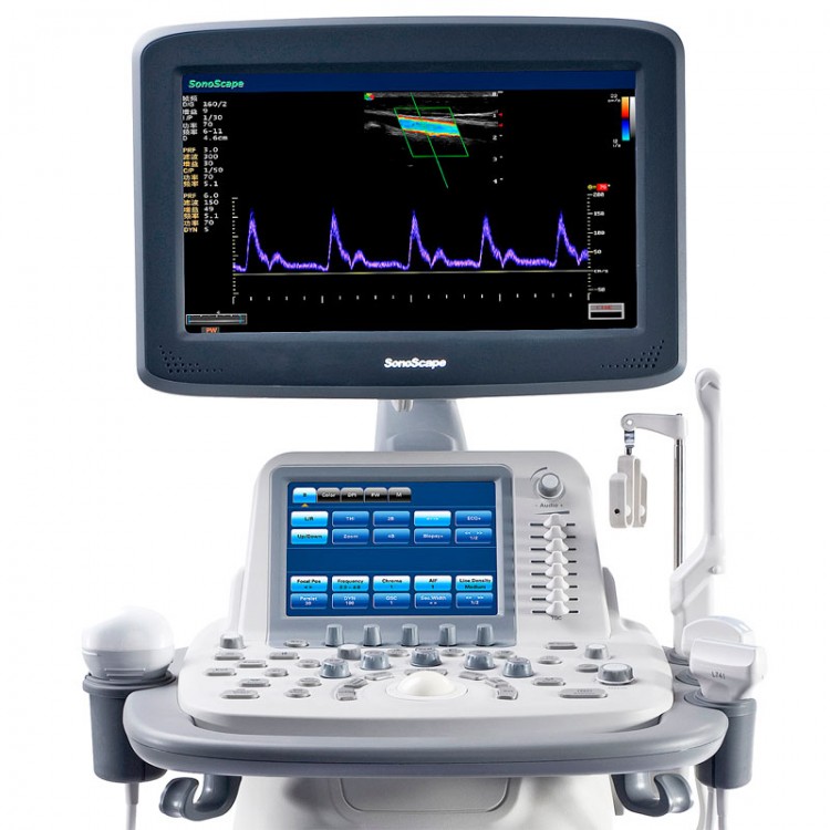
SonoScape S20Exp ultrasound machine

SonoScape S20Exp
The golden mean!
Appearance of S20Exp in the SonoScape (SonoScape) lineup means one thing – the new fast VIS platform becomes basic, replacing the former, perfectly proven S20 device.

The best price on the market for color ultrasound machines. Extremely short diagnostic times. All this is the key to the highest economic efficiency of SonoScape scanners.

Technical characteristics:
Large 17″ LCD monitor
4 active ports for connecting transducers, 8″ touch panel
Built-in battery, up to 120 minutes scanning time
Areas of use:
Abdominal Gynecology
Obstetrics
Urology
Thyroid
Breast Bone and muscle system
Cardiology
Pediatrics
Neurosonography
Invasive procedures
Vascular
Transcranial Studies
Scanning modes:
B, M, B/M, B/B, 4B, Tissue Harmonic
Biopsy needle navigation (biopsy guides), Biopsy needle enhanced visualization (illumination) mode
Real-time and still image scaling Color Doppler, Power Doppler, Power Doppler, Pulsed Wave Doppler (PW), HPRF, CW, Tissue Doppler (TDI)
Duplex, triplex modes
Compound Imaging (ultrasound beam swing mode)
Trapezoidal scanning on linear transducers
FreeHand 3D – surface 3D reconstruction mode
4D – mode of three-dimensional reconstruction in real time Anatomic M-mode, Color M-mode, Panoramic scan
Speckle-noise reduction technology MicroScan
Sonoelastography mode with quantitative assessment on linear, intracavitary and convex transducers (optional)
Ultrasonic contrast media mode on convex transducers
Digital workstation:
500 GB hard drive, USB 2.0, DVD-RW, Ethernet, DICOM 3.0
Calculations for obstetrics, gynecology, angiology, urology, pediatrics, superficial, abdominal organs, cardiology, ability to assess cardiovascular system, fetal brain, output of fetal growth curves, automatic analysis of intima-media complex thickness, semi-automatic calculation of ejection fraction by disc method (Simpson), semi-automatic calculation of collar space thickness
Drawing up and importing reports with the ability to add images
Maintaining a database of patients, the ability to save and search for images, clips, 3D-images in different fields of the database
SonoScape S20Exp ultrasound transducers
4-3C-A – 1-6MHz/R50mm, 70D, 128-element wideband convex ultrasound transducer
4-L743 – 4-15MHz/46mm, high frequency linear, high density ultrasonic transducer, 192 elements
4-6V3 – 4-11MHz/R10mm, 200D, microconvex rectovaginal, high-density ultrasonic transducer, 192 elements
4-2P1 – 1-5MHz, 90D, sector phased low frequency ultrasonic transducer

$2,213.00

Category:

Description

SonoScape S20Exp
The golden mean!
Appearance of S20Exp in the SonoScape (SonoScape) lineup means one thing – the new fast VIS platform becomes basic, replacing the former, perfectly proven S20 device.

The best price on the market for color ultrasound machines. Extremely short diagnostic times. All this is the key to the highest economic efficiency of SonoScape scanners.

Technical characteristics:
Large 17″ LCD monitor
4 active ports for connecting transducers, 8″ touch panel
Built-in battery, up to 120 minutes scanning time
Areas of use:
Abdominal Gynecology
Obstetrics
Urology
Thyroid
Breast Bone and muscle system
Cardiology
Pediatrics
Neurosonography
Invasive procedures
Vascular
Transcranial Studies
Scanning modes:
B, M, B/M, B/B, 4B, Tissue Harmonic
Biopsy needle navigation (biopsy guides), Biopsy needle enhanced visualization (illumination) mode
Real-time and still image scaling Color Doppler, Power Doppler, Power Doppler, Pulsed Wave Doppler (PW), HPRF, CW, Tissue Doppler (TDI)
Duplex, triplex modes
Compound Imaging (ultrasound beam swing mode)
Trapezoidal scanning on linear transducers
FreeHand 3D – surface 3D reconstruction mode
4D – mode of three-dimensional reconstruction in real time Anatomic M-mode, Color M-mode, Panoramic scan
Speckle-noise reduction technology MicroScan
Sonoelastography mode with quantitative assessment on linear, intracavitary and convex transducers (optional)
Ultrasonic contrast media mode on convex transducers
Digital workstation:
500 GB hard drive, USB 2.0, DVD-RW, Ethernet, DICOM 3.0
Calculations for obstetrics, gynecology, angiology, urology, pediatrics, superficial, abdominal organs, cardiology, ability to assess cardiovascular system, fetal brain, output of fetal growth curves, automatic analysis of intima-media complex thickness, semi-automatic calculation of ejection fraction by disc method (Simpson), semi-automatic calculation of collar space thickness
Drawing up and importing reports with the ability to add images
Maintaining a database of patients, the ability to save and search for images, clips, 3D-images in different fields of the database
SonoScape S20Exp ultrasound transducers
4-3C-A – 1-6MHz/R50mm, 70D, 128-element wideband convex ultrasound transducer
4-L743 – 4-15MHz/46mm, high frequency linear, high density ultrasonic transducer, 192 elements
4-6V3 – 4-11MHz/R10mm, 200D, microconvex rectovaginal, high-density ultrasonic transducer, 192 elements
4-2P1 – 1-5MHz, 90D, sector phased low frequency ultrasonic transducer

Reviews

There are no reviews yet.

Be the first to review “SonoScape S20Exp ultrasound machine”

Your email address will not be published. Required fields are marked *